
Spinal cord - Sekizui (English spelling)
|
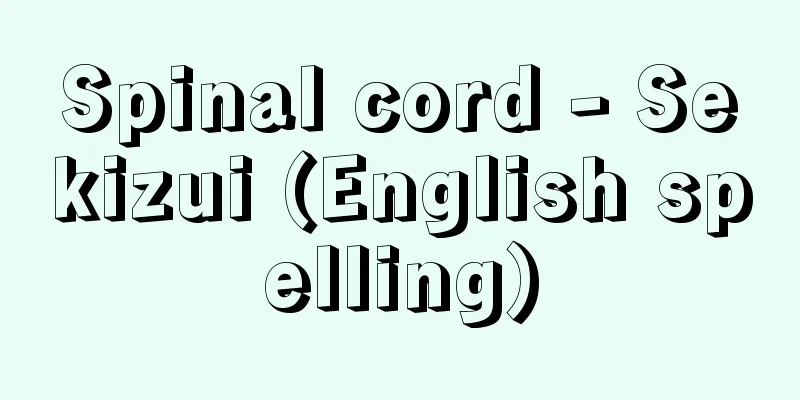
A white cord-like band that runs down the dorsal midline of vertebrates. Here, we will use humans as an example. The central nervous system is a single organ, but is divided into two parts, the brain and the spinal cord, based on its internal structure. Of these, the spinal cord is the part of the neural tube that is the basis for the formation of the brain and spinal cord that undergoes the least morphological change, and can be said to be the part where the primitive, somite-like arrangement remains intact. [Kazuyo Shimai] Location and FormThe spinal cord is connected to the lower part of the medulla oblongata at the bottom of the brain, and is located inside the spinal canal. It is elongated, approximately 1 cm in diameter, and has a white cylindrical shape that is flattened from front to back. The upper edge of the first cervical vertebra is where it meets the medulla oblongata. There are two slightly thicker parts along the way, called the cervical enlargement and the lumbar enlargement. The lower end of the spinal cord ends in a cone shape, and this part is called the conus medullaris. In adults, the conus medullaris is located at approximately the height of the first or second lumbar vertebra. At birth, the conus medullaris is located at the third lumbar vertebra, but it moves upward as the child grows. The length of the spinal cord is approximately 45 cm for Japanese adult males and 42-43 cm for Japanese adult females. A thin, thread-like fibrous tissue called the filum terminale descends from the lower end of the conus medullaris, reaching the position of the first coccyx and attaching to it. The filum terminale acts as an anchor to fix the spinal cord within the spinal canal. The spinal cord is surrounded and protected by three meninges (dura mater, arachnoid mater, and pia mater) that are the same as those of the brain. The spinal pia mater is the innermost layer and adheres closely to the surface of the spinal cord, but this pia mater protrudes triangular folds from both sides of the spinal cord, the tips of which penetrate the spinal arachnoid mater and attach to the spinal dura mater. These fold-like projections are called the dentate ligaments, and there are 18 to 20 pairs on the left and right, which serve to fix and support the spinal cord within the spinal canal. A deep longitudinal groove called the anterior median fissure runs along the anterior surface of the spinal cord, corresponding to the midline, and a shallow posterior median groove runs along the posterior surface, also corresponding to the midline. On either side of these grooves run the anterior lateral groove and the posterolateral groove, which run parallel to the two grooves, respectively, through which the spinal nerves enter and exit the spinal cord. The nerve bundle that passes through the anterior lateral groove is called the anterior root (motor root), and the nerve bundle that passes through the posterolateral groove is called the posterior root (sensory root). The anterior and posterior roots on the same side at the same height join just outside the spinal cord to form a single spinal nerve, which exits the spinal column through the intervertebral foramen between the vertebrae. In this way, a pair of spinal nerves emerge from the spinal cord at regular intervals. The spinal nerves are divided into 31 pairs in total: cervical (8 pairs), thoracic (12 pairs), lumbar (5 pairs), sacral (5 pairs), and coccygeal (1 pair). The part of the spinal cord where the cervical nerves emerge is called the cervical spinal cord, which is similarly divided into the thoracic, lumbar, and sacral spinal cord (including the coccygeal nerve). The dorsal root has a bulging area (called the spinal ganglion) just before where the dorsal root and anterior root join, and this area contains a group of nerve cell bodies that emerge from the nerve fibers in the dorsal root. [Kazuyo Shimai] Spinal cord nerve cellsThe internal structure of the spinal cord can be clearly seen when viewed in cross section. There is a thin vertical hole in the center, called the central canal. It continues to the fourth ventricle inside the medulla oblongata at the top, and ends at the conus medullaris at the bottom (called the ventricle terminalis). The central canal is filled with cerebrospinal fluid. The central canal is surrounded by a part called gray matter, which has an overall shape of a thick H. The outside of the gray matter is covered by white matter, and it appears white to the naked eye because it contains many myelinated nerve fibers. The H-shaped gray matter consists of two vertical lines on the left and right and a horizontal line connecting them, and the front part of the two lines is called the anterior horn and the rear part is called the posterior horn. The H-shaped gray matter forms an H-shaped gray column in three dimensions, so the anterior horn is called the anterior column and the rear horn is called the posterior column. In the lower cervical and upper thoracic spinal cord, the gray matter protrudes slightly to the side, which is called the lateral horn (forming a lateral column in three dimensions). The gray columns contain nerve cells, and the anterior horn is mainly composed of motor nerve cells, whose fibers form anterior roots and then exit the spinal cord to be distributed to the skeletal muscles of the entire body. The anterior horns of the cervical and lumbar enlargements contain well-developed nerve cells that send out motor nerves to the upper and lower limbs, respectively. It is generally believed that the cells on the inside of the anterior horn control the skeletal muscles of the trunk, and the cells on the outside control the skeletal muscles of the limbs. It is also believed that the cells that control the flexor muscles are arranged deep in the anterior horn, and the cells that control the extensor muscles are arranged shallowly in the anterior horn. On the other hand, the structure of the posterior horn is more complex than that of the anterior horn, but it is generally believed that it contains sensory nerve cells and secondary sensory cells that receive stimuli from primary sensory cells outside the spinal cord. Primary sensory cells are cell bodies in the spinal ganglia, and not only send fibers to various sensory receptors throughout the body, but also send fibers to the spinal cord through the dorsal roots to enter the posterior horn. In the spinal cord, sensory fibers form the dorsal roots and motor fibers form the ventral roots; this order is called Bell-Magendie's law (named after the Scottish physician Charles Bell (1774-1842) and the French physician François Magendie (1783-1855)). Sympathetic nerve cells exist in the lateral horn (lateral column), and their fibers pass through the anterior root and the sympathetic trunk to be distributed throughout the body. The white matter of the spinal cord is mainly composed of myelinated nerve fibers that run longitudinally. The part sandwiched between the anterior columns on both sides is called the anterior column, the part sandwiched between the anterior and posterior columns on the same side is called the lateral column, and the part sandwiched between the posterior columns on both sides is called the posterior column. Within this white matter, fibers with the following three physiological functions gather and run. The first type is the sensory nerve tract (ascending), which transmits excitation from each part of the body to the brain via the spinal cord. The second type is the motor nerve tract (descending), which transmits excitation from the brain to the skeletal muscles via the spinal cord. The third type is the nerve tract that connects various parts of the spinal cord. Of these, the motor nerve tract and the sensory nerve tract are called projection tracts because they mainly form long conduction paths, and the connecting nerve tracts within the spinal cord are called association tracts in the broad sense (note that there are association tracts in the narrow sense that connect the same side, and commissure tracts that connect both sides). There are four main sensory pathways: (1) The gracilis fasciculus and the cuneate fasciculus are nerve tracts that transmit discriminatory sensations (deep sensations) to the lower and upper body, respectively. (2) Anterior spinothalamic tract: A neural pathway that transmits non-discriminatory tactile sensations. (3) Lateral spinothalamic tract: A neural pathway that transmits pain and temperature sensations. (4) Anterior spinocerebellar tract and posterior spinocerebellar tract: Nerve pathways that connect the spinal cord and cerebellum. In addition, the following two are the main motor nerve tracts: (1) Pyramidal tract (corticospinal tract) A neural pathway that controls voluntary movement. It originates in the motor cortex of the cerebral cortex, descends to the spinal cord, and connects to the motor anterior horn cells. The pyramidal tract consists of the lateral corticospinal tract and the anterior corticospinal tract. (2) Extrapyramidal tracts These are motor nerve tracts related to movements other than those of the pyramidal tract. The main nerve tracts in this category are the tectalospinal tract, rubrospinal tract, vestibulospinal tract, reticulospinal tract, and medial longitudinal fasciculus. [Kazuyo Shimai] Spinal reflexThe spinal cord has a functionally important structure called spinal reflex. In other words, the spinal cord connects each part of the body to the brain and transmits a wide variety of signals, and in addition, it processes sensory information about environmental changes occurring inside and outside the body by involuntarily and unconsciously transmitting it to the motor cells of the spinal cord without passing through the cerebral cortex. This function is called spinal reflex. These spinal reflexes include stretch reflex and flexion reflex. The former is a reflex in which a skeletal muscle immediately responds and contracts when it is rapidly stretched. An example of this is the patellar tendon reflex. The patellar tendon reflex is when the patellar tendon is tapped, which stimulates the muscle to stretch, and the stimulation enters the posterior column of the spinal cord via the sensory nerves of the dorsal root, but part of the stimulation is transmitted to the anterior horn motor cells, causing the quadriceps muscle to contract and the lower leg to jump up. On the other hand, the flexor reflex is a reflex in which, for example, when the skin of a limb receives a painful stimulus, the flexor muscles of the entire limb contract, bending the limb to avoid the stimulus. In this way, spinal reflexes are highly integrated by the brain, but even if the brain were to be eliminated, the spinal cord alone would be able to function purposefully to a certain extent through its reflex circuits. In addition, the descending fibers of the autonomic nervous system descend from the autonomic center in the spinal cord, but no clear descending pathway is formed. The spinal cord is also thought to contain centers for autonomic reflexes such as sweating, vasomotion, piloerection, pupil dilation, respiratory movement, cardiac stimulation, lactation, and the urogenital system, but the details have not yet been elucidated. [Kazuyo Shimai] [Reference] | | |©Shogakukan "> Names of parts of the spinal cord ©Shogakukan "> An inside look at the spinal cord ©Shogakukan "> Cross-section of the spinal cord, major nerve pathways, and spinal reflexes Source: Shogakukan Encyclopedia Nipponica About Encyclopedia Nipponica Information | Legend |
|
脊椎(せきつい)動物の背側正中部を走る白色の索状帯。ここではヒトを例にして説明する。中枢神経は1個の器官であるが、その内部構造によって脳と脊髄という二つの部分に区分される。このうち脊髄は、脳脊髄形成の基である神経管のもっとも形態的変化が少ない部分で、いわば原始的な、体節的な配列がそのまま残っている部分といえる。 [嶋井和世] 位置と形態脊髄は、脳の最下部にある延髄の下方につながる部分で、脊柱管内に存在している。全体としては細長く、直径がほぼ1センチメートルの、前後に圧平された白い円柱状をしており、第1頸椎(けいつい)上縁あたりが延髄との境となる。途中の2か所にはやや太い部分があり、それぞれ頸膨大、腰膨大とよぶ。脊髄下端は円錐(えんすい)状に終わり、この部分を脊髄円錐とよぶ。成人の場合、脊髄円錐の位置は、ほぼ第1腰椎か第2腰椎の高さとなる。なお、出生時の脊髄円錐の位置は第3腰椎であるが、成長するにしたがって上方にあがっていく。脊髄の長さは日本人の成人男子では約45センチメートル、女子では42~43センチメートルとされる。脊髄円錐の下端からは糸状の細い終糸とよぶ線維組織が下降し、第1尾骨の位置まで達して、第1尾骨に付着する。終糸は、脊柱管内で脊髄を固定する錨(いかり)の役を果たしている。脊髄は脳と共通する3枚の髄膜(硬膜、クモ膜、軟膜)によって包まれ、保護されている。脊髄軟膜はいちばん内側にあって脊髄表面に密着しているが、この軟膜は脊髄両外側から三角状のヒダ(襞)を突出させ、その先端は脊髄クモ膜を貫いて脊髄硬膜に付着している。このヒダ状突起を歯状靭帯(じんたい)とよび、左右で18~20対あり、脊柱管内で脊髄の固定・支持の役を果たしている。 脊髄の前面には正中線に相当して前正中裂とよぶ深い縦溝が走り、後面には、やはり正中線に相当して浅い後正中溝が走る。両溝の両側外側には、それぞれ前外側溝、後外側溝が両溝と平行して走っているが、脊髄に出入りする脊髄神経はこの溝から出入りする。前外側溝を通り抜ける神経束を前根(運動根)とよび、後外側溝を通り抜ける神経束を後根(感覚根)とよぶ。同じ高さの同側の前根と後根とは、脊髄のすぐ外側で合流し、1本の脊髄神経となり、椎骨間の椎間孔を抜けて脊柱の外に出る。このようにして、脊髄神経は左右1対が一定間隔で脊髄から出ているわけである。 脊髄神経は、上から順に頸神経(8対)、胸神経(12対)、腰神経(5対)、仙骨神経(5対)、尾骨神経(1対)に区分し、合計31対となる。また、頸神経の出る脊髄部分を頸髄とよび、以下、同じように胸髄、腰髄、仙髄(尾骨神経を含む)に区分される。後根には、後根と前根とが合流するやや手前に膨らんだ部分があり(この部を脊髄神経節という)、ここには、後根内の神経線維を出す神経細胞体の集団が存在する。 [嶋井和世] 脊髄の神経細胞脊髄は横断面で見ると、その内部構造がよくわかる。中心部には細い縦孔があり、これを中心管とよぶ。上方は延髄内部の第四脳室に続き、下端は脊髄円錐の部分で終わる(この部を終室という)。この中心管内部には脳脊髄液が満たされている。中心管の周囲には灰白質という部分があり、全体形は太いH字形をしている。灰白質の外部を覆うのは白質で、肉眼で白くみえるのは多数の有髄神経線維を含むためである。H字形の灰白質は、左右の縦の2線と、これを結ぶ横線の部分とからなるが、2本線の前部を前角、後部を後角とよぶ。なお、H字形の灰白質は、立体的にはH字形の灰白柱となるため、前角は立体的には前柱、後角は後柱とよぶ。頸髄下部と胸髄上部では灰白質がわずかに側方に突出するので、これを側角とよぶ(立体的には側柱を形成する)。灰白柱には神経細胞が存在するが、前角には主として運動性の神経細胞が集まり、その線維は前根を形成したあと脊髄を出て、全身の骨格筋に分布する。頸膨大や腰膨大の前角には、それぞれ上肢、下肢に行く運動神経を出す神経細胞が発達しているが、このうち、一般には前角の内側にある細胞が体幹の骨格筋を、外側にある細胞が四肢の骨格筋を支配するとされている。また、屈筋支配の細胞は前角の深部に、伸筋支配の細胞は前角の浅部に配列するとされている。一方、後角の構造は前角に比べて複雑であるが、一般には感覚性の神経細胞が存在し、脊髄の外部にある第一次の感覚細胞からの刺激を受け取る第二次の感覚細胞が存在するとされる。第一次の感覚細胞は脊髄神経節にある細胞体であり、体中のさまざまな感覚受容器に線維を送るだけでなく、後根を通じて脊髄へと線維を送り後角に入る。脊髄において、感覚性線維は後根を形成し、運動性線維は前根を形成するが、この秩序をベル‐マジャンディの法則〔スコットランドの医学者Charles Bell(1774―1842)と、フランスの医学者François Magendie(1783―1855)にちなむ〕とよぶ。 側角(側柱)には交感性神経細胞が存在し、その線維は前根を通って、交感神経幹を経て、全身に分布する。脊髄白質の部分は、おもに縦走する有髄神経線維からなり、両側の前柱に挟まれた部分を前索、同側の前柱と後柱に挟まれた部分を側索、両側の後柱に挟まれた部分を後索とよんでいる。この白質の中では、次に述べる三つの生理的機能をもつ線維が集まって走行する。一つ目は身体各部からの興奮を脊髄を経て脳に伝える感覚性神経路(上行性)、二つ目は脳からの興奮を脊髄を経て骨格筋に伝える運動性神経路(下行性)、三つ目は脊髄の諸部分を連絡する神経路の3種類である。このうち、運動性神経路と感覚性神経路は、主として長い伝導路を形成するため投射路とよび、脊髄内の連絡神経路は広義の連合路とよぶ(なお、この連合路には同側を連絡する狭義の連合路と、両側を連絡する交連路とがある)。 感覚性神経路のおもなものは次の四つである。 (1)薄束(はくそく)と楔状束(けつじょうそく) それぞれ下半身、上半身の識別性の感覚(深部感覚)を伝える神経路。 (2)前脊髄視床路 非識別性の触圧覚を伝える神経路。 (3)外側脊髄視床路 痛覚と温度覚を伝える神経路。 (4)前脊髄小脳路と後脊髄小脳路 脊髄と小脳とを連絡する神経路。 また、運動性神経路では次の二つが主体となる。 (1)錐体路(皮質脊髄路) 随意運動をつかさどる神経路で、大脳皮質運動野からおこり、脊髄まで下行して運動性前角細胞に連絡する。錐体路は外側皮質脊髄路と前皮質脊髄路とからなる。 (2)錐体外路 錐体路以外の運動に関係する運動性神経路である。この神経路の主体をなすのは、視蓋(しがい)脊髄路、赤核脊髄路、前庭脊髄路、網様体脊髄路、内側縦束などである。 [嶋井和世] 脊髄反射脊髄には、脊髄反射という機能上重要となる構造がある。すなわち、脊髄は身体各部と脳との間を連絡し、多種多様の信号を伝えるほかに、体内・体外に生じる環境変化の感覚情報を、大脳皮質を経由せずに、不随意・無意識的に脊髄の運動細胞に伝えて感覚情報の処理にあたる。この働きをするのが脊髄反射である。この脊髄反射には伸張反射と屈曲反射とがある。前者は、骨格筋を急速に伸ばすと、その筋がただちに反応して収縮する反射であり、例として膝蓋腱(しつがいけん)反射があげられる。膝蓋腱反射とは、膝蓋腱をたたくとその刺激で筋が伸張し、その興奮は後根の感覚神経を経て脊髄後柱に入るが、興奮の一部は前角運動性細胞に伝えられ、大腿四頭筋(だいたいしとうきん)が収縮し、下腿が跳ね上がるという反射である。一方の屈曲反射とは、たとえば四肢の皮膚が痛みをおこすような刺激を受けたとき、屈筋が収縮し、肢全体の屈筋群が収縮して刺激を避けようとして四肢を曲げるような反射をいう。このように、脊髄反射は脳によって高次の統合を受けるが、かりに脳をなくしたとしても、脊髄だけである程度まではその反射回路によって、合目的的に働くことができるようになっている。 このほか、脊髄中には自律神経系の下行性線維が自律中枢から下行しているが、明瞭(めいりょう)な下行経路はつくられていない。また、脊髄には発汗、血管運動、立毛運動、瞳孔(どうこう)散大、呼吸運動、心臓促進、乳汁分泌、あるいは泌尿生殖系などの自律系反射の中枢もあると考えられているが、その細部についてはまだ判明していない。 [嶋井和世] [参照項目] | | |©Shogakukan"> 脊髄の各部名称 ©Shogakukan"> 脊髄の内部構造 ©Shogakukan"> 脊髄の横断面、おもな神経路、脊髄反射 出典 小学館 日本大百科全書(ニッポニカ)日本大百科全書(ニッポニカ)について 情報 | 凡例 |


Recommend
Oakamuro - Oakamuro
A marine fish of the genus Carangidae and family C...
Firm's product supply function
...To obtain the maximum profit possible, the rat...
Kineya Rokuzaemon
Nagauta shamisen player and singer. There have be...
Ihram - Ihram
… After the Battle of Badr in 624, Muhammad made ...
Evening crane
A play by Kinoshita Junji. One act. Published in ...
Nutritional enrichment - Eiyokyouka
…Therefore, the idea of supplementing foods wit...
Peroxysome
...Also called microbodies. Small granules in the...
Everyday life (English: vie quotidienne) (French)
It refers to the characteristics of steady, repet...
skepticism
… a word used as a translation of the Western phi...
Eccles, S. - Eccles
…English composer. The Eccles family was known as...
Documents in the womb - Tainai Monjo
Documents placed inside statues of Buddha and deit...
Halva (English spelling) Uno Harva
1882‐1949 A Finnish religious folklorist, he was a...
Immigrant
An individual or group moves from one country to ...
Common butterwort
A perennial plant of the Utricularia family (APG ...
Eireson Strait - Eireson Strait
…the name given to Sweden's dominance along t...