
Impulse conduction system
|
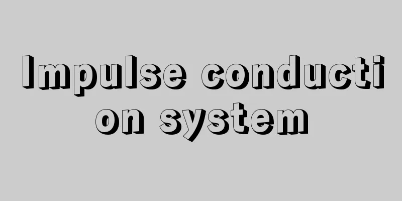
A specialized system of cardiac muscle cells (myocardial fibers) that controls the contraction of the heart. The cardiac muscle cells in this conduction system have the same function as normal cardiac muscle cells, but they are characterized by their function of only transmitting stimuli. The conduction system is composed of four structures: the sinoatrial node, the atrioventricular node, the atrioventricular bundle, and the Purkinje fibers. The sinoatrial node, also called the Keith-Flack node (named after physiologists A. Keith and M. Flack) or the pacemaker, is a small mass of tissue 2.5 cm long and 0.2 cm wide. This node is located near the opening of the superior vena cava in the wall of the right atrium, and a large number of cardiac muscle cells gather together to form a reticular structure. These cells have their own intrinsic contraction rhythm, so they do not require stimulation by nerve transmission from the brain or spinal cord. In other words, the muscle cells in the node themselves generate regular contraction stimuli, and the excitation stimuli are transmitted throughout the muscle layers of both atria. The excitation of this node is the beginning of the heartbeat, hence it is called a pacemaker or "pacing device." When the atrial muscle contracts due to the sinoatrial node, the impulse goes to the atrioventricular node. The atrioventricular node is also called the Tahara node (named after Jun Tahara (1873-1955), a professor of physiology at Kyushu University), and is also a small cluster of specialized cardiac muscle cells. The atrioventricular node is thicker than the sinoatrial node and is located on the posterior wall of the right atrium just above the opening of the coronary sinus. The excitation impulse of the atrioventricular node goes rapidly to the ventricles through the atrioventricular bundle. This atrioventricular bundle is also called the bundle of His (named after the internist W. His), and originates from the atrioventricular node, runs about 1-2 centimeters along the posterior-inferior border of the membranous part of the ventricular septum, and divides into the right and left bundle branches at the upper end of the muscular part of the ventricular septum. The right and left bundle branches descend toward the apex of the heart, just below the endocardium on the inner surface of the right and left ventricles, respectively, within the septum. Both bundle branches reach the bottom of the papillary muscles and are distributed in the muscular layer and papillary muscles of the right and left ventricles, respectively. The branches of the atrioventricular bundle are called Purkinje fibers (named after physiologist JE Purkinje). The atrial and ventricular muscular layers are completely cut off at the boundary of the annulus fibrosus, but this conduction system is the only one that connects the atrial and ventricular muscles. These specialized cells are thicker than ordinary cardiac muscle cells, rich in myocyte characteristics, and have few myofibers. Excitation can occur from any part of the conduction system, but the sinoatrial node has the highest frequency of excitation, so the heart generally functions with the sinoatrial node as a pacemaker, as mentioned above. When the atrioventricular bundle is blocked, the contraction order of the atria and ventricles is disrupted, and they contract independently. This condition is called atrioventricular block. [Kazuyo Shimai] [Reference] |©Shogakukan "> Components of the Conduction System Source: Shogakukan Encyclopedia Nipponica About Encyclopedia Nipponica Information | Legend |
|
心臓の収縮運動をつかさどる特殊な心筋細胞(心筋線維)系をいう。この伝導系の心筋細胞群は、収縮という機能に関しては普通の心筋細胞と同じであるが、刺激伝達だけに働くというのが特徴である。刺激伝導系は四つの構造、すなわち洞房結節、房室結節、房室束、プルキンエ線維から構成されている。洞房結節はキース‐フラック結節(生理学者A. KeithとM. Flackの名にちなむ)、あるいはペースメーカーともよばれ、長さ2.5センチメートル、幅0.2センチメートルの小組織塊である。この結節は右心房の壁の上大静脈の開口近くに存在し、多数の心筋細胞が集まって網状構造をつくっている。これらの細胞は本来、固有の収縮リズムをもっているため、脳や脊髄(せきずい)からの神経伝達による刺激は必要としない。つまり、結節の筋細胞自身で規則的な収縮刺激を生じ、その興奮刺激は両側の心房の筋層の至る所に伝わるわけである。この結節の興奮が心臓拍動の始まりとなるために、ペースメーカー、あるいは「歩調とり」とよばれるわけである。洞房結節によって心房筋が収縮すると、その刺激は房室結節へ進む。房室結節は田原結節〔田原淳(1873―1955)九州大学生理学教授の名にちなむ〕ともよばれ、やはり特殊な心筋細胞の小塊である。房室結節は洞房結節よりも太く、右心房の後壁で冠状静脈洞の開口のすぐ上に存在する。房室結節の興奮刺激は房室束を通って急速に心室に進む。この房室束はヒス束(内科学者W. Hisの名にちなむ)ともよばれ、房室結節からおこり、心室中隔の膜性部の後下縁に沿って約1~2センチメートル走り、心室中隔筋性部の上端で右脚と左脚とに分かれる。右脚と左脚とはそれぞれ中隔の中で右室と左室の内面の心内膜直下を心尖(しんせん)に向かって下降する。両脚は乳頭筋の底部に到達し、それぞれ右室と左室の筋層や乳頭筋に分布する。房室束の分枝をプルキンエ線維(生理学者J. E. Purkinjeにちなむ)とよんでいる。心房筋層と心室筋層とは線維輪を境にして完全に連絡を絶たれているが、この伝導系だけが心房筋と心室筋との間を連ねている。この特殊細胞は一般の心筋細胞よりも太く、筋細胞形質にも富み、筋細線維が少ないのが特徴である。刺激伝導系ではどの部分からでも興奮がおこりうるが、洞房結節の興奮頻度がもっとも大きいため、一般には前述したように洞房結節をペースメーカーとして心臓機能が発揮されている。なお、房室束が遮断されると、心房と心室の収縮秩序が乱されて、それぞれがばらばらに収縮する状態となる。この状態を房室ブロックという。 [嶋井和世] [参照項目] |©Shogakukan"> 刺激伝導系の構成要素 出典 小学館 日本大百科全書(ニッポニカ)日本大百科全書(ニッポニカ)について 情報 | 凡例 |
<<: Shigeno story - Shige Shigeyawa


Recommend
Equalizer
1. Something that equalizes. Something that brings...
lovat mixture
…(5) Lovat This is a relatively new fabric, creat...
diner (English spelling)
…the most important meal of the day, or a meal wi...
《Otsuma Hachirobei》 - Otsuma Hachirobei
...Sewamono (domestic drama). Also called "T...
Thalassemia
Hemolytic anemia caused by a genetic defect and ba...
Mando-e
A Buddhist ceremony in which 10,000 lanterns or a...
Carneades's board - Carneades's board
It is one of the ethical thought experiments, but...
Photometer
...The term photometer is generally used, but thi...
Puṣyamitra (English spelling) Puṣamitra
…An ancient North Indian dynasty (c. 180-68 BC). ...
Ezo starfish - Ezohitode
It is a marine animal belonging to the family Sta...
Not matching
…Pumice deposits often weather to a yellowish bro...
Battle of Pydna - Battle of Pydna
A war fought in 168 B.C. in Pydna, on the Aegean c...
Intoku Taiheiki - Intoku Taiheiki
A military chronicle set in Western Japan during t...
Nobuharu Baba
1513‐75 (Eisho 10‐Tensho 3) A military commander o...
Cartaphilus
…Christ then replied, “Wait for me to come,” and ...